Geometry
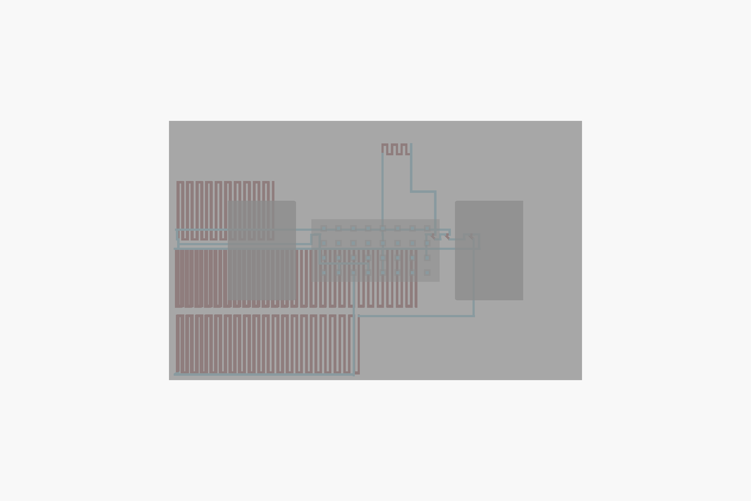
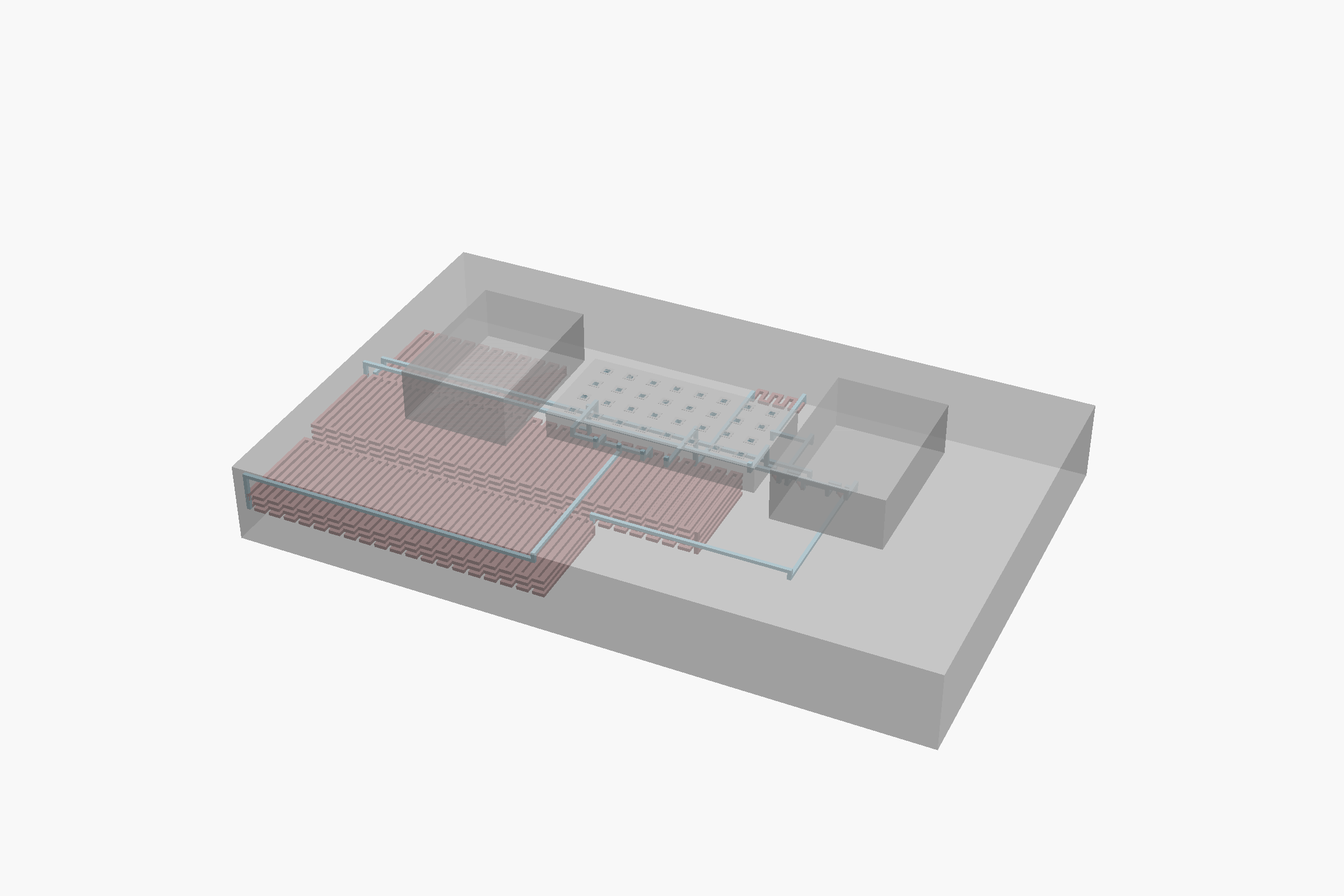
Top view (left) and isometric view (right) of the placed-and-routed chip. Rendered with OpenSCAD from the chip’s SCAD source, albumin_reroute.scad. Click for high-resolution.
Download: PNG (top) · PNG (iso) · SCAD source ↓
Specifications
Assay
This chip is designed to measure urinary albumin concentration via an immunoturbidimetric assay. Anti-human-albumin antibodies bind albumin in the sample and form aggregates that scatter light; the resulting turbidity is measured spectrophotometrically and is proportional to the albumin concentration in the original sample.
Clinically, urinary albumin quantification supports kidney function screening: albumin in urine (microalbuminuria) is an early marker of glomerular damage, with strong predictive value for chronic kidney disease in diabetic and hypertensive patients.
The chemistry is adapted from the Beckman Coulter AU480 reagent kit BSB38858 (Microalbumin). The chip implements passive resistance-metering to combine the urine sample with three reagent streams at the volumetric ratios required by the assay protocol; the output channel collects the mixed product for off-chip spectrophotometric readout.
Files
- SCAD
albumin_reroute.scad1.7 MBDownload - LEF
h.r.3.3_albumin_pcells.lef2.4 KBDownload - CSV
pcell_out_scad266 BDownload - PNG
albumin_iso_large.png119.5 KBDownload - PNG
albumin_top_large.png19.6 KBDownload
albumin_reroute.scad is the placed-and-routed 3D geometry — the main OpenSCAD source for this chip. h.r.3.3_albumin_pcells.lef contains chip-specific cell abstracts. pcell_out_scad lists the parametric serpentine-channel variants this chip required (with L1 / L2 / turn count). The PNG renders are high-resolution versions of the views shown in the Geometry section above.
Papers
-
Related methodology archetype
Goenner, B., Temple, S., Zapata, S., Wakeham, D., Snelgrove, A., Gaillardon, P.-E., Nordin, G. P., & Gale, B. K. (2025). An open source platform to automate the design, verification, and manufacture of 3D printed microfluidic devices. Scientific Reports, 15, 33077. DOI: 10.1038/s41598-025-15976-9
Applications
For research use only. Not for use in diagnostic procedures.
A design for kidney function screening by urinary albumin quantification: albumin in urine is an early marker of glomerular damage and chronic kidney disease. One of the urinalysis chip designs in the Gale Lab urinalysis chip family (Erica Francis).